
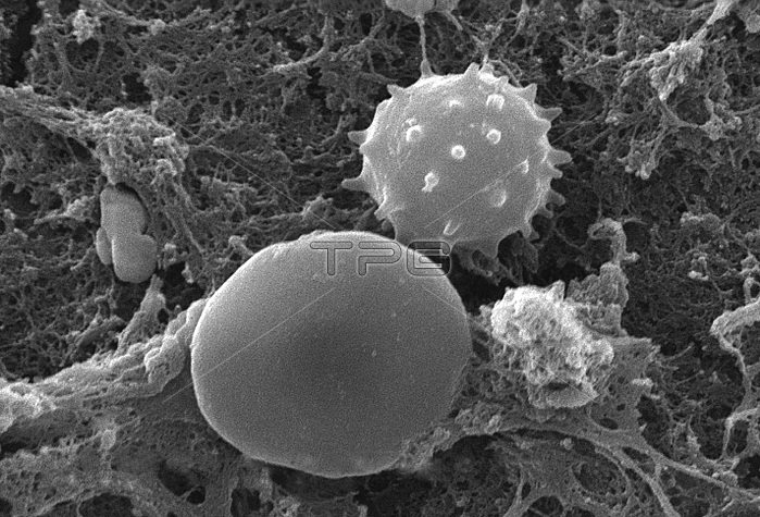
This scanning electron micrograph (SEM) depicted a number of red blood cells found enmeshed in a fibrinous matrix on the luminal surface of an indwelling vascular catheter; Magnified 7766x. In this instance, the indwelling catheter was a tube that was left in place creating a patent portal directly into a blood vessel. The cell in the center was a white blood cell, also known as a leucocyte. The biconcave cytomorphologic shape of the red blood cell, or erythrocyte, increases its surface area of this hemoglobin-filled cell, thereby, promoting a greater degree of gas exchange, which is its primary function in an in vivo setting. In their adult phase, these cells possess no nucleus. What appears to be irregularly-shaped chunks of debris, are actually fibrin clumps, which when inside the living organism, functions as a key component in the process of blood clot formation, acting to entrap the red blood cells in a mesh-like latticework of proteinaceous strands, thereby, stabilizing and strengthening the clot, in much the same way as rebar acts to strengthen, and reinforce cement.
| px | px | dpi | = | cm | x | cm | = | MB |
Details
Creative#:
TOP22228218
Source:
達志影像
Authorization Type:
RM
Release Information:
須由TPG 完整授權
Model Release:
N/A
Property Release:
No
Right to Privacy:
No
Same folder images:
 Loading