
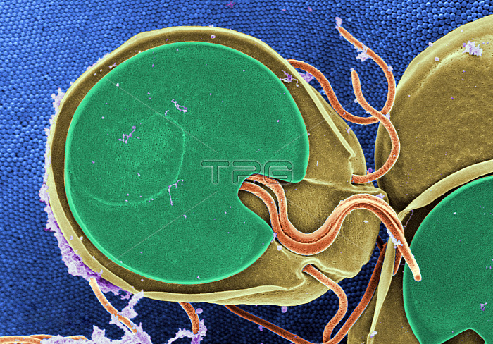
Color-enhanced Scanning Electron Micrograph (SEM) showing the ventral surface of a Giardia muris trophozoite that had settled atop the mucosal surface of a rat's intestine. Note the microvilli, which can be seen in the background, as tiny rounded structures that are approximately 0.15 microns in diameter. These microvilli cover the surface of each intestinal epithelial cell. The Giardia's ventral adhesive disk resembles a suction cup, where overlapping microtubules in the cytoplasm form a number-6-shaped figure. The edge of the suction cup, called the ventrolateral flange, partially encircles the adhesive disk and is absent posteriorly where a ventral pair of flagella emerges from above, dorsal to the disk. Giardia muris has four pairs of flagella that are responsible for the organism's motility. The adhesive disk facilitates adherence to the intestinal surface.
| px | px | dpi | = | cm | x | cm | = | MB |
Details
Creative#:
TOP22228453
Source:
達志影像
Authorization Type:
RM
Release Information:
須由TPG 完整授權
Model Release:
N/A
Property Release:
No
Right to Privacy:
No
Same folder images:
 Loading