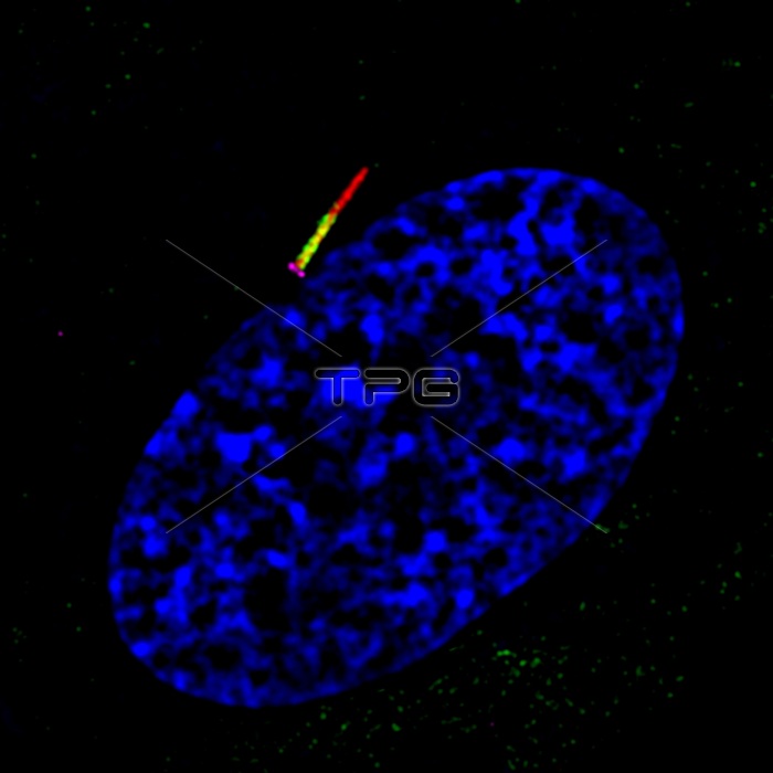
Understanding the mechanisms regulating transport of signaling molecules to and from the cilia is important for developing drugs targeted against ciliary-associated cancer pathways. The ciliary membrane is continuous with the plasma membrane through a specialized pocket-like membrane referred to as the ciliary pocket. Here, structured illumination microscopy (SIM) imaging of RPE cells shows the ciliary membrane and ciliary pocket. The membrane reshaping protein EHD1 (green) specifically accumulates at the ciliary pocket membrane while the Hedgehog pathways receptor Smoothened localizes to the ciliary membrane (red). The base of the cilia is marked by the mother centriole distal appendage protein CEP164 (pink) and nuclei (blue).
| px | px | dpi | = | cm | x | cm | = | MB |
Details
Creative#:
TOP22235321
Source:
達志影像
Authorization Type:
RM
Release Information:
須由TPG 完整授權
Model Release:
N/A
Property Release:
No
Right to Privacy:
No
Same folder images:

 Loading