
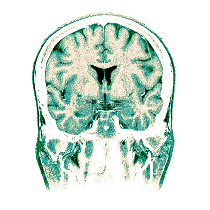
This is a normal coronal (frontal view) cross-sectional MRI image of the brain through both frontal lobes and both temporal lobes. At this level you see two cavities filled with dark material near the center of the image. These are the lateral ventricles which are filled with cerebral spinal fluid (CSF). There are two main types of brain tissue, gray matter (which contains the neuronal cell bodies and is the darker of the brain tissue shown) and white matter (which is composed of axonal fibers).
| px | px | dpi | = | cm | x | cm | = | MB |
Details
Creative#:
TOP22289787
Source:
達志影像
Authorization Type:
RM
Release Information:
須由TPG 完整授權
Model Release:
N/A
Property Release:
No
Right to Privacy:
No
Same folder images:
 Loading