
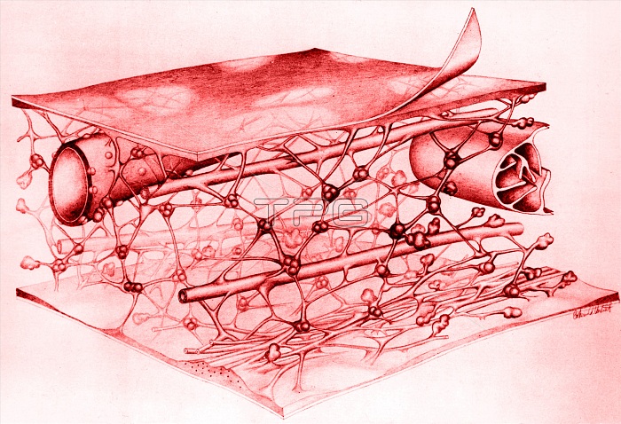
Color enhanced schematic of the microtrabecular lattice in the cytoplasm of a thinly spread cell in culture as seen with the high voltage electron microscope. The trabeculae are joined at thicker nodal points. The lattice is traversed by microtubules and the trabeculae appear to attach to these, to mitochondria, and to the cell membrane.
| px | px | dpi | = | cm | x | cm | = | MB |
Details
Creative#:
TOP22292410
Source:
達志影像
Authorization Type:
RM
Release Information:
須由TPG 完整授權
Model Release:
N/A
Property Release:
No
Right to Privacy:
No
Same folder images:
 Loading