
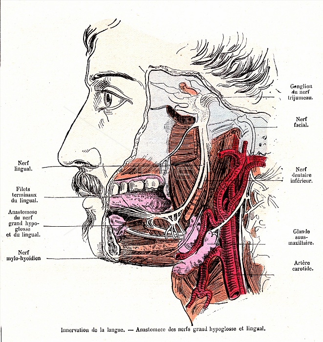
Tongue and mouth nerve anatomy, 19th-century illustration. The nerves shown here branch out from upper right. The facial nerve (down right) is shown with a branch supplying the submaxillary salivary gland (pink), with the nerve shown passing in front of the carotid artery. At upper centre, the trigeminal nerve ganglion is shown. Also known as the fifth cranial nerve (CN V), this nerve and its branches produce sensation in the face, as well as functions such as biting and chewing. Its three major branches are the ophthalmic nerve (not shown), the maxillary nerve (partially shown) and the mandibular nerve (shown here). The latter branches into several nerves, including the lingual nerve (to the tongue), the inferior alveolar nerve (also called the inferior dental nerve) and the mylohyoid nerve. Also shown in the tongue is an area of connection (anastomosis) between the lingual nerve and the hypoglossal nerve. The labels are in French. Published in 'La Vie Normale et la Sante' (Normal Life and Health, Paris, 1881) by Dr Jules Rengade.
| px | px | dpi | = | cm | x | cm | = | MB |
Details
Creative#:
TOP24751673
Source:
達志影像
Authorization Type:
RM
Release Information:
須由TPG 完整授權
Model Release:
N/A
Property Release:
N/A
Right to Privacy:
No
Same folder images:
 Loading