
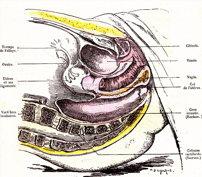
Female pelvic organs anatomy, 19th-century illustration. This sagittal section through a woman's pelvic region shows (from top): urinary system organs, reproductive system organs, and the final parts of the digestive system. Organs shown here and labelled include the bladder (upper centre), the uterus (below and left of the bladder) and the rectum (lower centre). Other structures and body parts include the clitoris, the fallopian tubes, the ovaries, the vagina, the cervix (neck of the uterus). The final part of the backbone is across bottom, and include the sacrum and coccyx. The labels are in French. Published in 'La Vie Normale et la Sante' (Normal Life and Health, Paris, 1881) by Dr Jules Rengade.
| px | px | dpi | = | cm | x | cm | = | MB |
Details
Creative#:
TOP24751676
Source:
達志影像
Authorization Type:
RM
Release Information:
須由TPG 完整授權
Model Release:
N/A
Property Release:
N/A
Right to Privacy:
No
Same folder images:
 Loading