
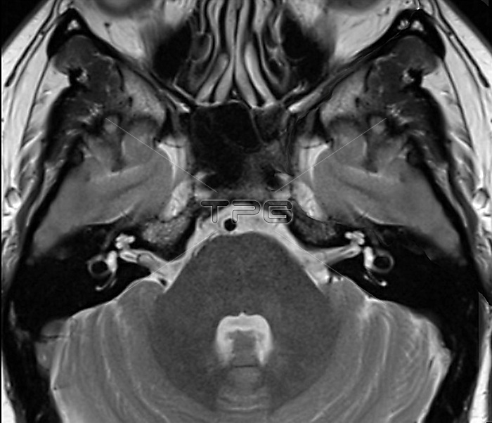
Axial section of the brain and inner ear. T2-weighted magnetic resonance imaging (MRI) scan of an axial section of the brain. The scan is centred on the subtentorial space (region under the cerebella tentorium containing the pons, medulla oblongata, cerebellum and mesencephalon). This is the region allowing for hearing and balance. The bony labyrinth (left and right) is the rigid outer wall of the inner ear, and is made up of the cochlea, the semicircular canals, and the vestibule which connects the former two parts. Internal auditory ducts connect this region to the vestibulocochlear nerve (eighth cranial nerve), which allows the transmission of sound and equilibrium (balance) information from the inner ear to the brain.
| px | px | dpi | = | cm | x | cm | = | MB |
Details
Creative#:
TOP24873032
Source:
達志影像
Authorization Type:
RM
Release Information:
須由TPG 完整授權
Model Release:
N/A
Property Release:
N/A
Right to Privacy:
No
Same folder images:
 Loading