
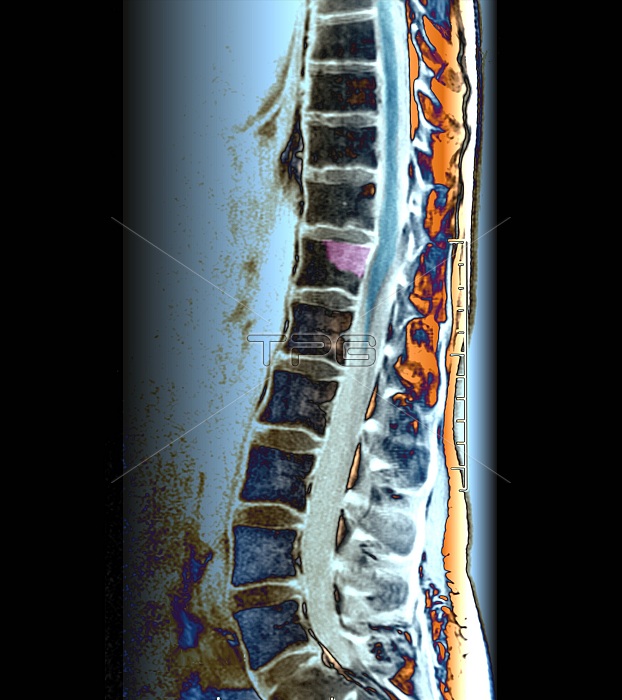
Secondary bone cancer in the spine. Magnetic resonance imaging (MRI) scan of a medial sagittal section of the thoracolumbar spine of a 32-year-old female patient with a primary melanoma (cancer) of the tibia (shinbone). There is evidence of secondary (metastatic) tumours in the bones of the spine (vertebrae), specifically the T12 vertebra and the tissue surrounding it. There is also evidence that the cancer has infiltrated the spinal chord. Primary bone cancer is very rare, however cancerous metastatic spread forming secondary bone tumours (specifically in the vertebrae) is fairly common. The primary melanoma of the tibia would likely be treated through an above-knee amputation to remove the section of cancerous bone, or alternatively chemotherapy or radiotherapy to destroy the cancerous cells. This is a T1-weighted MRI scan.
| px | px | dpi | = | cm | x | cm | = | MB |
Details
Creative#:
TOP24873056
Source:
達志影像
Authorization Type:
RM
Release Information:
須由TPG 完整授權
Model Release:
N/A
Property Release:
N/A
Right to Privacy:
No
Same folder images:
 Loading