
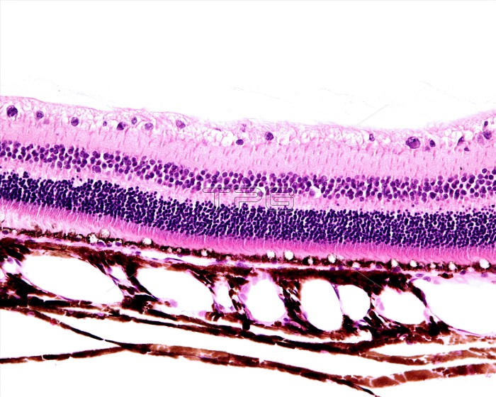
Light micrograph showing the retina and choroid. From top to bottom, the retina layers can be seen: nerve fibre layer, ganglion cell layer, inner plexiform layer, inner nuclear layer, outer plexiform layer, outer nuclear layer, rods and cones layer, and pigment epithelium layer. Outside the pigment epithelium, the pigmented choroid shows dilated blood vessels.
| px | px | dpi | = | cm | x | cm | = | MB |
Details
Creative#:
TOP25373473
Source:
達志影像
Authorization Type:
RM
Release Information:
須由TPG 完整授權
Model Release:
N/A
Property Release:
N/A
Right to Privacy:
No
Same folder images:
histologylightmicroscopenobodyno-onemicrographmicroscopemicroscopicmicroscopyhistologybiologyeyeeyeballophthalmologyophthalmicocularretinapigmentedepitheliumphotoreceptornervefibrelayerganglioncelllayerinnerplexiformlayerinnernuclearlayerouterplexiformlayerouternuclearlayerrodsconeshistologicallightmicrographlmbiological
biologicalbiologycellconesepitheliumeyeeyeballfibreganglionhistologicalhistologyhistologyinnerinnerlayerlayerlayerlayerlayerlayerlightlightlmmicrographmicrographmicroscopemicroscopemicroscopicmicroscopynerveno-onenobodynuclearnuclearocularophthalmicophthalmologyouterouterphotoreceptorpigmentedplexiformplexiformretinarods
 Loading