
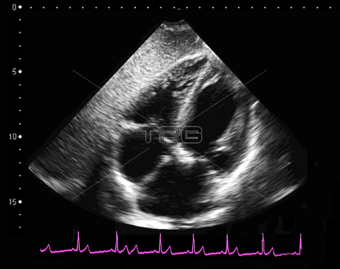
Normal heart. Ultrasound scan of a healthy heart in a 49-year-old man during the systolic phase of the heartbeat. This scan passes vertically through the heart, with the heart's four chambers seen as the black areas. The central white areas are the walls and valves separating the chambers. The top of the heart is at bottom. The top two chambers are the ventricles, and the bottom two chambers are the atria. The heart is a hollow muscular pump that contracts continuously under a steady cycle of electrical impulses, sending blood to the lungs and around the body. An electrocardiogram (ECG) trace is seen across bottom, with the peaks of electrical activity corresponding to the beating of the heart. Ultrasound scanning uses high-frequency sound waves to look at structures inside the body.
| px | px | dpi | = | cm | x | cm | = | MB |
Details
Creative#:
TOP25477221
Source:
達志影像
Authorization Type:
RM
Release Information:
須由TPG 完整授權
Model Release:
N/A
Property Release:
N/A
Right to Privacy:
No
Same folder images:
 Loading