
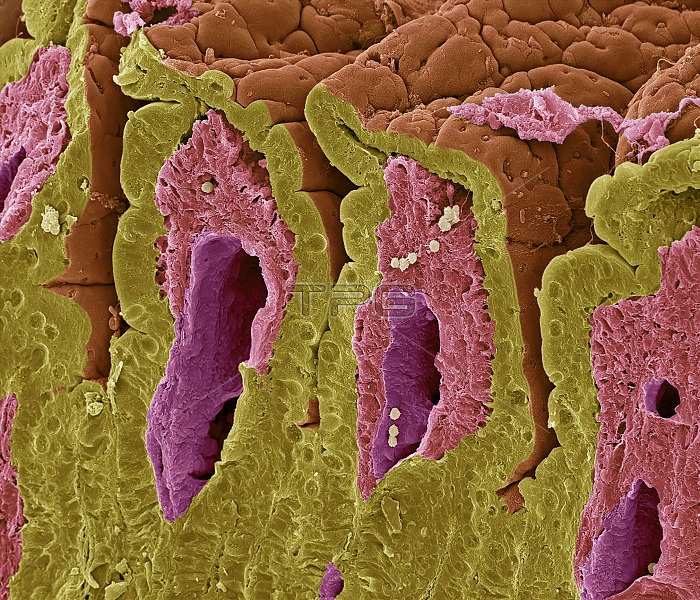
Intestinal lining. Coloured scanning electron micrograph (SEM) of a freeze-fractured surface of the small intestine. The surface consists of deep folds, called villi, that have been sectioned in this view. The intestinal surface (orange, upper frame) is exposed to food. The underlying structure of this surface is seen in the sectioned area. Surface (epithelial) cells (yellow) are supported by connective tissue (pink- purple) that forms the core of each fold (villus). This central core of connective tissue is known as the lamina propria. This contains large blood vessels, capillaries, some smooth muscle cells and a blind- ended lymph vessel known as a lacteal. The folds increase the area for the absorption of nutrients from food. The height of a villus varies in a small intestine from 0.3 to 0.8 millimetres. Magnification: x800 when printed at 10 centimetres wide.
| px | px | dpi | = | cm | x | cm | = | MB |
Details
Creative#:
TOP25487583
Source:
達志影像
Authorization Type:
RM
Release Information:
須由TPG 完整授權
Model Release:
N/A
Property Release:
N/A
Right to Privacy:
No
Same folder images:
 Loading