
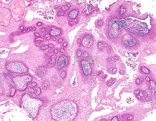
Craniopharyngioma, light micrograph. Craniopharyngioma is a燱HO Grade 1 epithelial tumour爑sually seen in the爏ellar/suprasellar region. It arises most likely from the爎emnants of Rathke's pouch. Some cases may have their origin in爉isplaced odontogenic rests along pituitary stalk. Grossly, they are often爏olid and cystic with areas of calcification. Craniopharyngiomas are subdivided into two types based on morphologic features: 1) Adamantinomatous (90% of cases) seen mostly in children and 2) Papillary (10% of cases) seen mostly in adults. Papillary craniopharyngiomas more often tend to be solid and rarely show calcification (unlike adamantinomatous variant). They are composed of sheets of well-differentiated non-keratinizing squamous epithelium (WHO Grade 1). Dehiscence of epithelium around fibrovascular cores often results in pseudopapillary architecture as seen here.
| px | px | dpi | = | cm | x | cm | = | MB |
Details
Creative#:
TOP25929026
Source:
達志影像
Authorization Type:
RM
Release Information:
須由TPG 完整授權
Model Release:
N/A
Property Release:
N/A
Right to Privacy:
No
Same folder images:
 Loading