
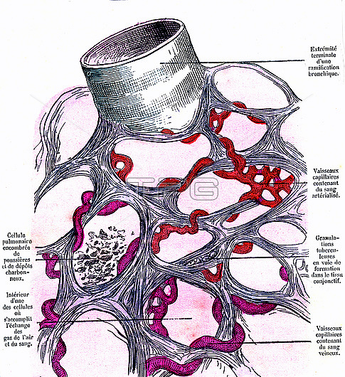
Lung alveoli anatomy, 19th-century illustration. At upper left is part of a terminal bronchiole, which ends in the alveolar sacs shown below in cross-section. Venous (purple) and arterial (red) capillaries are shown meshed in the alveoli, which are the site of gas exchange in the lungs. Carbon dioxide and oxygen are the gases exchanged here. Also shown are deposits of carbon (lower left), and tuberculosis deposits (tubercules, centre). The labels are in French. Published in 'La Vie Normale et la Sante' (Normal Life and Health, Paris, 1881) by Dr Jules Rengade.
| px | px | dpi | = | cm | x | cm | = | MB |
Details
Creative#:
TOP26513881
Source:
達志影像
Authorization Type:
RM
Release Information:
須由TPG 完整授權
Model Release:
N/A
Property Release:
N/A
Right to Privacy:
No
Same folder images:
1800sabnormalairwaysalveolianatomicalbiologicalbloodvesselscapillariescapillarybedcarbonconditiondisordereuropeanfrenchfrenchlanguagegasexchangehealthyhistoricaljulesrengadelavienormaleetlasantelabellabeledlabelledlabelslungsmedicalno-onenobodynormalnormallifeandhealthparticulateparticulatespollutionpulmonaryrespiratorysoottbtexttuberculestuberculosisunhealthyvascularorgandiseaseairwayalveolushumanbodychestlunganatomybiologyhistorymedicinepneumologyartworkillustration19thcentury1881
1800s188119thabnormalairwayairwaysalveolialveolusanatomicalanatomyandartworkbedbiologicalbiologybloodbodycapillariescapillarycarboncenturychestconditiondiseasedisordereteuropeanexchangefrenchfrenchgashealthhealthyhistoricalhistoryhumanillustrationjuleslalalabellabeledlabelledlabelslanguagelifelunglungsmedicalmedicineno-onenobodynormalnormalnormaleorganparticulateparticulatespneumologypollutionpulmonaryrengaderespiratorysantesoottbtexttuberculestuberculosisunhealthyvascularvesselsvie
 Loading