
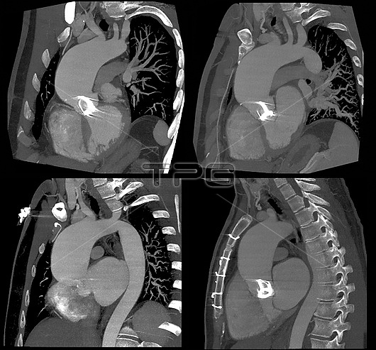
Thoracic aortic aneurysm. Computed tomography angiogram (CTA) scans of sections through the heart of a 41-year-old patient with a thoracic aortic aneurysm. At age 20, this patient had an operation for aortic valve stenosis (narrowing). The aortic valve lies between the left ventricle of the heart and the aorta (main artery carrying oxygenated blood from the heart). The patient now has a thoracic aortic aneurysm (dilatation) of the upper aorta caused by the weakening of the arterial wall. If an aneurysm bursts it can cause severe internal haemorrhage (bleeding). Here the aneurysm measures 60mm across the major aortic axis and extends to the brachiocephalic artery (largest branch of the aortic arch that supplies blood to the head, neck, and right arm). The aneurysm also measures 37mm at the sinus of Valsalva (a small pouch that is directly above the aortic valve). There is also a narrowing of the aortic isthmus (smaller branch of the aortic arch).
| px | px | dpi | = | cm | x | cm | = | MB |
Details
Creative#:
TOP26514880
Source:
達志影像
Authorization Type:
RM
Release Information:
須由TPG 完整授權
Model Release:
N/A
Property Release:
N/A
Right to Privacy:
No
Same folder images:
 Loading