
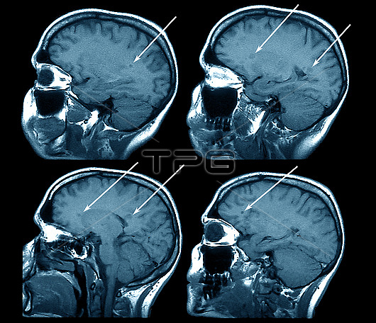
Multiple sclerosis. Coloured magnetic resonance imaging (MRI) scans of left parasagittal sections of the brain of a 37-year-old patient with multiple sclerosis (MS). MS is due to the destruction of the protective myelin sheaths surrounding the axon nerve fibres of the brain and spinal cord. Several demyelinated lesions (damaged areas, indicated by arrows) can be seen in the white matter of the patient's brain. Axons in the affected area can no longer conduct nerve impulses, resulting in symptoms ranging from tingling to paralysis. This is caused by neuronal degradation and a gradual loss of nerve function. MS is believed to be an autoimmune disorder in which the immune system attacks myelin. This is a T1-weighted MRI scan.
| px | px | dpi | = | cm | x | cm | = | MB |
Details
Creative#:
TOP26514914
Source:
達志影像
Authorization Type:
RM
Release Information:
須由TPG 完整授權
Model Release:
N/A
Property Release:
N/A
Right to Privacy:
No
Same folder images:
 Loading