
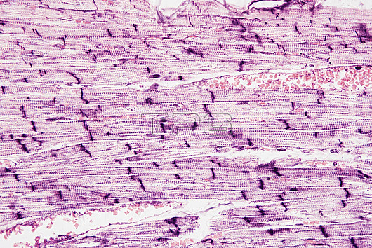
Light micrograph of a longitudinal section through cardiac (heart) muscle. This section is from a papillary muscle of the heart. Cardiac muscle consists of branching elongated muscle cells, called myocytes, which form fibres (running horizontally). The myocytes are separated by intercalated discs (purple vertical lines), which allow electrical signals to travel rapidly between myocytes. Myocytes consist of microfilaments of the contractile proteins actin and myosin and a centrally located nucleus. Cardiac muscle is myogenic, it can cause its own contraction without any other input. Magnification: x400 when printed at 15cem wide.
| px | px | dpi | = | cm | x | cm | = | MB |
Details
Creative#:
TOP27148448
Source:
達志影像
Authorization Type:
RM
Release Information:
須由TPG 完整授權
Model Release:
N/A
Property Release:
N/A
Right to Privacy:
No
Same folder images:
 Loading