
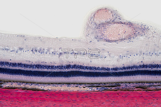
Light micrograph of a section through the eye of the wall showing the retina. The sclera (red), the white of the eye is at bottom. Above this is the choroid, which contains blood vessels. Above this is the retina whose first layer is a line of pigment cells (black), the retinal pigment epithelium, that lines the inside of the eye and prevents light from reflecting and distorting the image. This is immediately followed by the light sensitive rod and cone cells (light pink). For light to reach these cells it must pass through layers of nerve cells. The nerve cell nuclei are visible as the layers of purple and blue dots. Two blood vessels are at top left. Magnification: x200 when printed at 15cm wide.
| px | px | dpi | = | cm | x | cm | = | MB |
Details
Creative#:
TOP27148466
Source:
達志影像
Authorization Type:
RM
Release Information:
須由TPG 完整授權
Model Release:
N/A
Property Release:
N/A
Right to Privacy:
No
Same folder images:
 Loading