
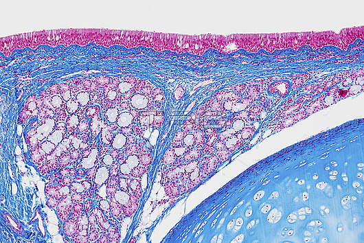
Light micrograph of a section through the trachea, or windpipe, the tube leading from the mouth to the bronchus and the lungs. It is formed by a number of horseshoe-shaped rings of cartilage, cut crosswise here and seen as the blue area at bottom right. At the top are the columnar epithelial cells (pink) that line the trachea. They have hair-like cilia on their surface that beat to move mucus, and particles trapped in it, upwards out of the respiratory tract. The mucus is produced by mucosa glands (pink, hollow) and secreted by goblet cells in the epithelium. Magnification: x100 when printed at 15cm wide.
| px | px | dpi | = | cm | x | cm | = | MB |
Details
Creative#:
TOP27148525
Source:
達志影像
Authorization Type:
RM
Release Information:
須由TPG 完整授權
Model Release:
N/A
Property Release:
N/A
Right to Privacy:
No
Same folder images:
 Loading