
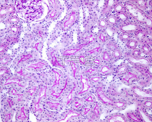
Light micrograph of a renal cortex stained with the periodic acid Schiff (PAS) method. The PAS method allows distinction between the proximal convoluted tubules (most of the tubules seen here) and the distal convoluted tubules. Proximal convoluted tubules have a PAS positive brush border. Distal convoluted tubules have a broader lumen, lower epithelium and are devoid of a brush border. Here the distal tubules are sparse, but some can be seen in the vicinity of the glomerulus at the upper edge.
| px | px | dpi | = | cm | x | cm | = | MB |
Details
Creative#:
TOP27562919
Source:
達志影像
Authorization Type:
RM
Release Information:
須由TPG 完整授權
Model Release:
N/A
Property Release:
N/A
Right to Privacy:
No
Same folder images:
 Loading