
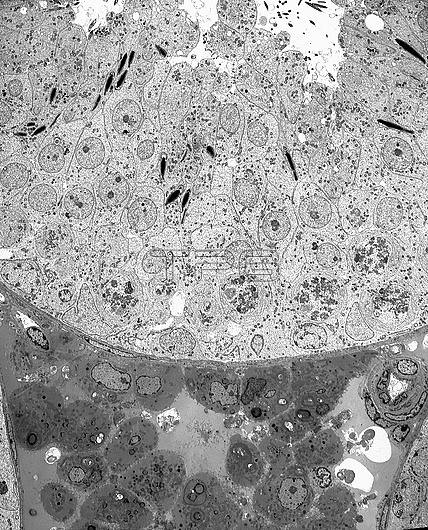
Transmission electron micrograph (TEM) of the ultrastructure of the seminiferous epithelium and intertubular tissue of the testis. In the epithelium, Sertoli cells and spermatogonia are located at the base with primary spermatocytes and round and elongating spermatids positioned above and towards the seminiferous tubule lumen. Numerous Leydig cells and a blood vessel are seen in the intertubular tissue. Magnification: x750 when height printed at 10cm.
| px | px | dpi | = | cm | x | cm | = | MB |
Details
Creative#:
TOP27779081
Source:
達志影像
Authorization Type:
RM
Release Information:
須由TPG 完整授權
Model Release:
N/A
Property Release:
N/A
Right to Privacy:
No
Same folder images:
TestisseminiferousepitheliumseminiferoustubulespermatogenesisSertolicellspermatogoniumspermatocyteprimaryspermatocytespermatidintertubulartissueLeydigcellcellbiologyultrastructurecellultrastructureelectronmicrographelectronmicroscopytransmissionelectronmicrographmalemalereproductionmalereproductivesystemtemnobodyno-oneblackandwhitemonochromebiologicalcytologycytological
LeydigSertoliTestisandbiologicalbiologyblackcellcellcellcellcytologicalcytologyelectronelectronelectronepitheliumintertubularmalemalemalemicrographmicrographmicroscopymonochromeno-onenobodyprimaryreproductionreproductiveseminiferousseminiferousspermatidspermatocytespermatocytespermatogenesisspermatogoniumsystemtemtissuetransmissiontubuleultrastructureultrastructurewhite
 Loading