
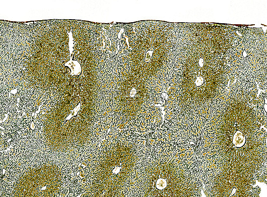
Light micrograph of liver tissues stained to show the connective tissue framework that supports the liver lobules. This connective tissue is referred to as reticular fibres made of type III collagen. Lobules form polygonal cords of hepatocytes surrounding a central vein seen here as empty holes. In disease conditions such as cirrhosis and hepatitis the connective tissue of collagen is produced in excess to form much fibrosis. This causes resistance to blood flow and compromises liver function. Paraffin section, Gordon and Sweet's reticulin stain. Magnification: x20 when width printed at 10cm.
| px | px | dpi | = | cm | x | cm | = | MB |
Details
Creative#:
TOP27954921
Source:
達志影像
Authorization Type:
RM
Release Information:
須由TPG 完整授權
Model Release:
N/A
Property Release:
No
Right to Privacy:
No
Same folder images:
 Loading