
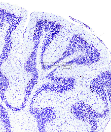
Light micrograph of a sagittal section of a cerebellum stained with cresyl violet. The cerebellar cortex is tightly folded in many branched cerebellar folia which resembles a tree ('tree of life'). In each folium can be seen three layers: the molecular layer, granular layer and central axis of white matter. Purkinje cells are located between the molecular and granular layer.
| px | px | dpi | = | cm | x | cm | = | MB |
Details
Creative#:
TOP28014702
Source:
達志影像
Authorization Type:
RM
Release Information:
須由TPG 完整授權
Model Release:
N/A
Property Release:
N/A
Right to Privacy:
No
Same folder images:
 Loading