
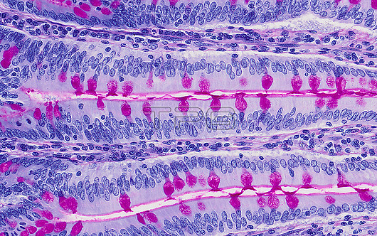
Small intestine, light micrograph. Small intestine villi have a core of connective tissue forming a lamina propria supporting a simple columnar epithelium. Nerve fibres, smooth muscle, vessels and lymphoid cells are found in the lamina propria. The absorption of nutrients occurs across the many enterocytes (blue). Secretion of mucus by the small bowel is provided by the goblet cells (red) with secreted mucus seen as a thin layer which coexists with epithelial surface microvilli. Together this layer is known as the 'brush border' as the microvilli (beyond the optical resolution seen here) resemble the hairs of a brush. Paraffin section, haematoxylin eosin/PAS stain. Magnification: x225 when width printed at 10cm.
| px | px | dpi | = | cm | x | cm | = | MB |
Details
Creative#:
TOP28014779
Source:
達志影像
Authorization Type:
RM
Release Information:
須由TPG 完整授權
Model Release:
N/A
Property Release:
N/A
Right to Privacy:
No
Same folder images:
 Loading