
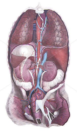
Posterior view of the thoracic and abdominal viscera, illustration. Between the lungs is the thoracic aorta (red), azygos vein (blue) and thoracic duct (white). Below the cut diaphragm at left is the stomach (white) and spleen (deep red) and to the right is the vena cava (blue) and the liver (red). Renal arteries supply the kidneys from which the ureters (white) descend inferiorly. The abdominal aorta and inferior vena cava in the pelvic region are associated with their respective iliac vessels. The ascending colon (white) lies below the right kidney with the sigmoid colon (white) below the left kidney. From Lizars, J. 1823 A system of anatomical plates of the human body. W.H. Lizars, Edinburgh.
| px | px | dpi | = | cm | x | cm | = | MB |
Details
Creative#:
TOP28328611
Source:
達志影像
Authorization Type:
RM
Release Information:
須由TPG 完整授權
Model Release:
N/A
Property Release:
N/A
Right to Privacy:
No
Same folder images:
 Loading