
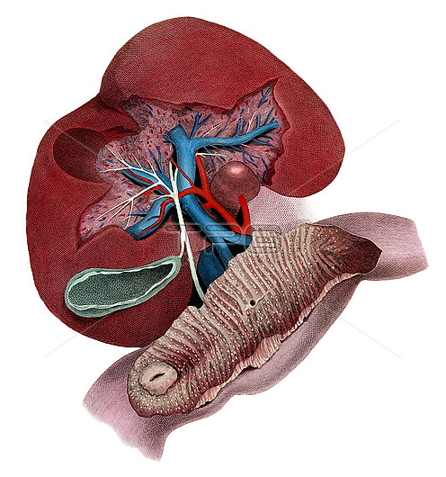
Illustration of a dissection of the liver to show the anatomy of the porta hepatis. The confluence of the superior mesenteric and splenic veins forms the portal vein (blue) that enters the liver with major branching. The hepatic artery (red) and gastric artery (cut) are shown. Bile from the liver passes into the hepatic ducts that join with the cystic duct from the gall bladder (green) to become the common bile duct that drains into the duodenum via an orifice at the duodenal papilla. The duodenum has been opened. To the left is the opening of the pyloric canal. Ridges of duodenal mucosa are termed plicae circulares. From Lizars, J. 1823 A system of anatomical plates of the human body. W.H. Lizars, Edinburgh.
| px | px | dpi | = | cm | x | cm | = | MB |
Details
Creative#:
TOP28328615
Source:
達志影像
Authorization Type:
RM
Release Information:
須由TPG 完整授權
Model Release:
N/A
Property Release:
N/A
Right to Privacy:
No
Same folder images:
 Loading