
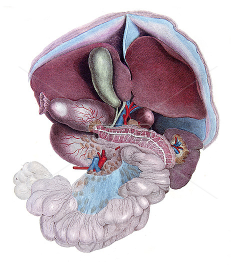
Abdominal viscera displayed after reflection of the liver. Uppermost is the cut diaphragm and the peritoneum with suspensory ligaments (blue) of the liver which has left and right lobes. Below the gall bladder (green) is the common bile duct (green), portal vein (blue) and hepatic artery (red). To the left the cut stomach leads to the duodenum into which the pancreatic duct (white) drains. The spleen and its vessels are located to the right of the tail of the pancreas. Vessels in the abdomen are the superior mesenteric artery (red) and vein (blue), below which is the flat sheet of mesentery (blue) attached to the loops of small bowel (jejunum). From Lizars, J. 1823 A system of anatomical plates of the human body. W.H. Lizars, Edinburgh.
| px | px | dpi | = | cm | x | cm | = | MB |
Details
Creative#:
TOP28328622
Source:
達志影像
Authorization Type:
RM
Release Information:
須由TPG 完整授權
Model Release:
N/A
Property Release:
N/A
Right to Privacy:
No
Same folder images:
 Loading