
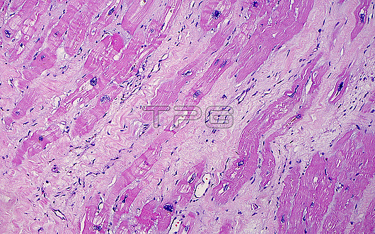
Light micrograph of heart muscle showing hypertrophy. Hypertrophy occurs when the heart muscle is under stress as occurs by increased demands on a heart weakened by lack of ischemia (lack of blood flow) or other disease. Microscopically, hypertrophy is seen as an increase in size of the heart muscle nuclei (the dark blue structures). There is also fibrosis (light pink areas) in between the heart muscle cells (darker pink areas), which is also the result of weakness and chronic damage of the heart muscle. Haematoxylin and eosin stained tissue section. Magnification: 100x when printed at 10 cm.
| px | px | dpi | = | cm | x | cm | = | MB |
Details
Creative#:
TOP28634953
Source:
達志影像
Authorization Type:
RM
Release Information:
須由TPG 完整授權
Model Release:
n/a
Property Release:
n/a
Right to Privacy:
No
Same folder images:
 Loading