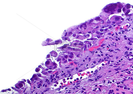
Light micrograph of in situ urothelial carcinoma (CIS), which is a premalignant condition or one that has the potential to develop into invasive cancer. CIS is composed enlarged, hyperchromatic (dark staining), and irregular shaped cells. The picture shows cells of CIS spanning left bottom to right top of the image. The right upper to lower half of the image shows the benign connective tissue underneath the abnormal CIS cells. Haematoxylin and eosin stained tissue section. Magnification: 200x when printed at 10 cm.
| px | px | dpi | = | cm | x | cm | = | MB |
Details
Creative#:
TOP28634955
Source:
達志影像
Authorization Type:
RM
Release Information:
須由TPG 完整授權
Model Release:
n/a
Property Release:
n/a
Right to Privacy:
No
Same folder images:
pathologydiseasedisorderconditiondiagnosisdiagnosticmedicinegenitourinarypathologydiseasedisorderconditiondiagnosisdiagnostichistologyhistologicalhistopathologydiseasedisorderconditiondiagnosisdiagnosticmicroscopylmmagnifiedimagelightmicrographslidehumanbodyanatomytissuecellsH&EstainHaematoxylinandeosinurinarysystembladderbladdercanceroncologyurologydiseaseabnormalwhitebackground
H&EHaematoxylinabnormalanatomyandbackgroundbladderbladderbodycancercellsconditionconditionconditiondiagnosisdiagnosisdiagnosisdiagnosticdiagnosticdiagnosticdiseasediseasediseasediseasedisorderdisorderdisordereosingenitourinaryhistologicalhistologyhistopathologyhumanimagelightlmmagnifiedmedicinemicrographmicroscopyoncologypathologypathologyslidestainsystemtissueurinaryurologywhite

 Loading