
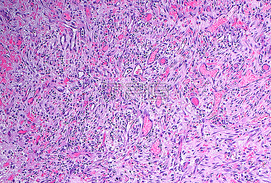
Light micrograph of granulation tissue. Granulation tissue forms when inflammation has been present in tissues for days to a few weeks. Granulation tissue is made up of small blood vessels (pink tubular or circular structures) mixed with inflammatory cells (small blue dots) in a myxoid stroma (light blue background throughout). Haematoxylin and eosin stained tissue section. Magnification: 100x when printed at 10 cm.
| px | px | dpi | = | cm | x | cm | = | MB |
Details
Creative#:
TOP28634964
Source:
達志影像
Authorization Type:
RM
Release Information:
須由TPG 完整授權
Model Release:
n/a
Property Release:
n/a
Right to Privacy:
No
Same folder images:
 Loading