
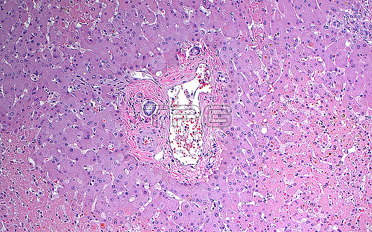
Light micrograph of a centrilobular pattern of necrotic liver cells. The necrotic liver cells are the lighter pink cells mostly in the bottom left and bottom right corners of the picture. In the centre of the image is a portal tract consisting of an artery, vein, and bile duct. The liver cells around the portal tract are not necrotic because they receive an adequate supply of blood from the nearby vessels. The liver cells further out are necrotic, indicating ischemia or a lack of enough oxygen and nutrients further away from the blood source. Such a pattern of necrosis can occur when there are other factors in the body that decrease the availability of oxygen or nutrients in the blood, such as infection or shock. Haematoxylin and eosin stained tissue section. Magnification: 100x when printed at 10 cm.
| px | px | dpi | = | cm | x | cm | = | MB |
Details
Creative#:
TOP28634981
Source:
達志影像
Authorization Type:
RM
Release Information:
須由TPG 完整授權
Model Release:
n/a
Property Release:
n/a
Right to Privacy:
No
Same folder images:
 Loading