
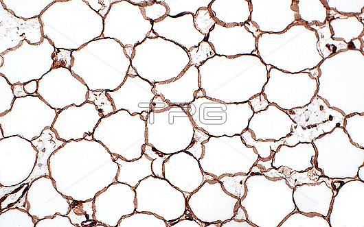
Light micrograph of adipocytes (fat cells) stained by immunohistochemistry, a technique where antibodies detect specific proteins in tissue by attaching to them and giving off a certain colour. Here, the antibody used detected the protein vimentin, a protein commonly present in most cell types. The brown colour of the stain highlights the membranes of the fat cells. The fat cells themselves are mostly made up of the white spaces which is where lipids (fat) is normally stored. Haematoxylin and eosin stained tissue section. Magnification: 200x when printed at 10cm.
| px | px | dpi | = | cm | x | cm | = | MB |
Details
Creative#:
TOP28635086
Source:
達志影像
Authorization Type:
RM
Release Information:
須由TPG 完整授權
Model Release:
n/a
Property Release:
n/a
Right to Privacy:
No
Same folder images:
 Loading