
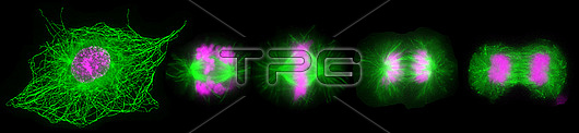
Fluorescent light micrograph of cells during mitosis (nuclear division). Mitosis is the formation of two daughter nuclei from one parent nucleus. Fluorescent markers have been used to highlight DNA (deoxyribonucleic acid, pink) and alpha tubulin (green), a component of microtubules. The cell at left is in prophase, the nuclear envelope is dissolving and the chromosomes are condensing. The cell progress through prometaphase (second from left) to metaphase (centre), where the chromosomes align along the centre of the cell. The chromosomes start to move to the opposite poles, guided by microtubules, during anaphase (second from right). The cell at right is in telophase, when the separated chromosomes have moved to opposite ends of the cell and two new nuclei form around them.
| px | px | dpi | = | cm | x | cm | = | MB |
Details
Creative#:
TOP28927411
Source:
達志影像
Authorization Type:
RM
Release Information:
須由TPG 完整授權
Model Release:
n/a
Property Release:
n/a
Right to Privacy:
No
Same folder images:
 Loading