
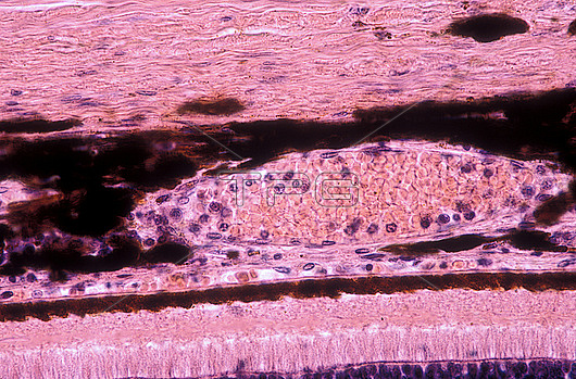
Light micrograph showing the choroid and retina. From top to bottom are the: sclera, heavily pigmented choroid with a large blood vessel (Haller?檚 layer) and small capillaries next to the retina (choriocapillaris), and outermost layers of retina, i.e. pigment epithelium, rod and cones layer. Near the bottom is the external limiting layer and some nuclei of the outer nuclear layer.
| px | px | dpi | = | cm | x | cm | = | MB |
Details
Creative#:
TOP28927727
Source:
達志影像
Authorization Type:
RM
Release Information:
須由TPG 完整授權
Model Release:
n/a
Property Release:
n/a
Right to Privacy:
No
Same folder images:
 Loading