
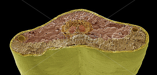
Pine needle. Coloured scanning electron micrograph (SEM) of a freeze-fracture through a leaf (needle) of a pine tree (Pinus sp.). The leaves are needle-like in order to present a large surface area for photosynthesis but prevent too much water loss (transpiration). They have an epidermis of thick walled cells covered with a thick layer of cuticle. The mesophyll layer under the epidermis is made up of parenchyma cells. The vascular cylinder (centre) is surrounded by the endodermis which regulates water and mineral movement. The centre of the needle is occupied by two vascular bundles, each one made up of xylem and phloem tissue. These are surrounded by a thick pericycle (large-celled region) and a layer of endodermis (necklace-like ring of large cells). A resin duct is seen at either side of the section. Magnification: x46 when printed 10 centimetres wide.
| px | px | dpi | = | cm | x | cm | = | MB |
Details
Creative#:
TOP28946831
Source:
達志影像
Authorization Type:
RM
Release Information:
須由TPG 完整授權
Model Release:
N/A
Property Release:
N/A
Right to Privacy:
No
Same folder images:
 Loading