
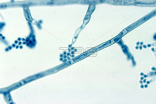
Light micrograph of the fungus Madurella mycetomatis showing conidiophores (branches with terminal clusters of ovals) and septate hyphae (one diagonally across centre). Hyphae are the branching, filamentous structures of a fungus, that in this species are separated by septa (walls) into cellular compartments. Conidiophores are specialised hyphae that produce asexual spores (reproductive cells, ovals) called conidia. M. mycetomatis can cause an infection of the subcutaneous tissue (deepest layer of skin) known as mycetoma.
| px | px | dpi | = | cm | x | cm | = | MB |
Details
Creative#:
TOP28948735
Source:
達志影像
Authorization Type:
RM
Release Information:
須由TPG 完整授權
Model Release:
n/a
Property Release:
n/a
Right to Privacy:
No
Same folder images:
asexualreproductionbiologicalbiologyconidiaconidiogenesisconidiophoreconidiumfilamentousfilamentsfungalfungifungushyphahyphaehyphallightmicrographlmmadurellamycetomatismicro-organismmicrobiologicalmicrobiologymicrographmicroorganismmicroscopymyceliamyceliummycologicalmycologyno-onenobodypathogenpathogenicphialidesseptatehyphaseptatehyphaesporewhitebackground
asexualbackgroundbiologicalbiologyconidiaconidiogenesisconidiophoreconidiumfilamentousfilamentsfungalfungifungushyphahyphahyphaehyphaehyphallightlmmadurellamicro-organismmicrobiologicalmicrobiologymicrographmicrographmicroorganismmicroscopymyceliamyceliummycetomatismycologicalmycologyno-onenobodypathogenpathogenicphialidesreproductionseptateseptatesporewhite
 Loading