
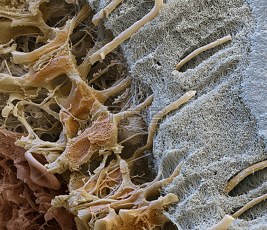
Coloured scanning electron micrograph (SEM) of a freeze-fracture through a human tooth. At far right is the dentine (blue), the mineralised connective tissue that forms the bulk of the tooth. To its left is a dense network of collagen known as predentine (grey), which will calcify into dentine. Within the dentine are dentine tubules, which have been formed by the cytoplasmic extensions (beige) of odontoblast cells (dentine-producing cells). The odontoblast cells originate in the pulp (red, bottom left) and allow the dentine to rebuild itself. The picture was created as part of a cooperation with Quintessenz Publishing. Magnification: x4,500 when printed at 15cm wide.
| px | px | dpi | = | cm | x | cm | = | MB |
Details
Creative#:
TOP28976564
Source:
達志影像
Authorization Type:
RM
Release Information:
須由TPG 完整授權
Model Release:
Not Available
Property Release:
Not Available
Right to Privacy:
No
Same folder images:
Restriction:
All moral rights asserted. Recolouring or alterationof the image is prohibited without permission.
 Loading