
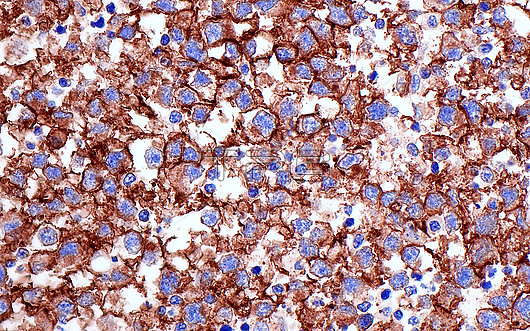
Light micrograph of cells of a seminoma, a type of testicular cancer, stained with the c-kit protein using an immunohistochemical technique. The tumour cells have blue stained nuclei (light blue circles) and the c-kit protein is detected by the brown colour of the stain. Magnification: x400 when printed at 10cm wide.
| px | px | dpi | = | cm | x | cm | = | MB |
Details
Creative#:
TOP29066904
Source:
達志影像
Authorization Type:
RM
Release Information:
須由TPG 完整授權
Model Release:
Not Available
Property Release:
Not Available
Right to Privacy:
No
Same folder images:
pathologymedicinepathologicalmedicalanatomicpathologytesticularcancerreproductivesystemtestistestistumourtumortumourcellsgenitourinarypathologyurologyoncologybiologyhistologyhistologicalhistopathologymicroscopylmlightmicrographslideimmunohistochemicalstainnobodyno-onehumanbodyanatomytissuecellswhitebackgroundabstract
abstractanatomicanatomybackgroundbiologybodycancercellscellsgenitourinaryhistologicalhistologyhistopathologyhumanimmunohistochemicallightlmmedicalmedicinemicrographmicroscopyno-onenobodyoncologypathologicalpathologypathologypathologyreproductiveslidestainsystemtesticulartestistestistissuetumortumourtumoururologywhite
 Loading