
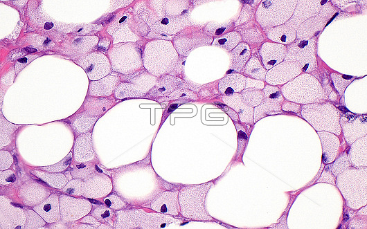
Light micrograph of necrotic (dead) fat cells. The fat cells (round white circles) are surrounded by foamy histiocytes (smaller light grey-pink circles), which are a type of chronic inflammatory cell. Fat necrosis occurs when the fat cells are damaged due to trauma or other disease processes. Haematoxylin and eosin stained tissue section. Magnification: x400 when printed at 10cm wide.
| px | px | dpi | = | cm | x | cm | = | MB |
Details
Creative#:
TOP29066932
Source:
達志影像
Authorization Type:
RM
Release Information:
須由TPG 完整授權
Model Release:
Not Available
Property Release:
Not Available
Right to Privacy:
No
Same folder images:
 Loading