
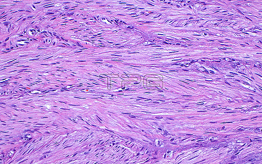
Light micrograph of cells of a uterine leiomyoma (fibroid), or a benign (non-cancerous) smooth muscle tumour growth. The cells of the leiomyoma have spindled nuclei (short and thin dark blue lines) and cytoplasm (pink) that forms intersecting fascicles or fibres together with other cells. Haematoxylin and eosin stained section. Magnification: x200 when printed at 10cm wide.
| px | px | dpi | = | cm | x | cm | = | MB |
Details
Creative#:
TOP29066957
Source:
達志影像
Authorization Type:
RM
Release Information:
須由TPG 完整授權
Model Release:
Not Available
Property Release:
Not Available
Right to Privacy:
No
Same folder images:
pathologymedicinepathologicalmedicalanatomicpathologyuterusleiomyomabenignfibroidsmoothmuscletumourtumorneoplasmgynaecologygynaecologicpathologyreproductivesystemfemalereproductivesystembiologyhistologyhistologicalhistopathologymicroscopylmlightmicrographslideHematoxylinHaematoxylinandeosinstainnobodyno-onehumanbodyanatomytissuecellsabstract
HaematoxylinHematoxylinabstractanatomicanatomyandbenignbiologybodycellseosinfemalefibroidgynaecologicgynaecologyhistologicalhistologyhistopathologyhumanleiomyomalightlmmedicalmedicinemicrographmicroscopymuscleneoplasmno-onenobodypathologicalpathologypathologypathologyreproductivereproductiveslidesmoothstainsystemsystemtissuetumortumouruterus
 Loading