
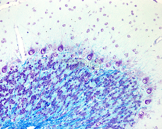
Light micrograph of cerebellar cortex stained with Luxol fast blue and cresyl violet. Myelinated fibres of the white matter, stained in blue with Luxol, can be seen in the white matter (bottom) and in the granular layer. The cresyl violet shows the Nissl bodies in Purkinje cells and three Golgi cells located in the granular layer.
| px | px | dpi | = | cm | x | cm | = | MB |
Details
Creative#:
TOP29699459
Source:
達志影像
Authorization Type:
RM
Release Information:
須由TPG 完整授權
Model Release:
N/A
Property Release:
N/A
Right to Privacy:
No
Same folder images:
cerebellumcnsbiologycerebellarcortexgranularlayerpurkinjegolgicellsfibrefibrefoliumgranularlayergreymattercresylviolethistologicalhistologylightmicroscopemicrographmicroscopicalmicroscopymolecularlayermoleculemyelinatednervenervousneurohistologyneurologicalneurologyluxolfastbluecresylvioletnobodyno-onelmlightmicrographmicroscopyhistologyhistologicalbiologybiologicalnormalhealthy
biologicalbiologybiologybluecellscerebellarcerebellumcnscortexcresylcresylfastfibrefibrefoliumgolgigranulargranulargreyhealthyhistologicalhistologicalhistologyhistologylayerlayerlayerlightlightlmluxolmattermicrographmicrographmicroscopemicroscopicalmicroscopymicroscopymolecularmoleculemyelinatednervenervousneurohistologyneurologicalneurologyno-onenobodynormalpurkinjevioletviolet
 Loading