- Share
- Download
Caption: 
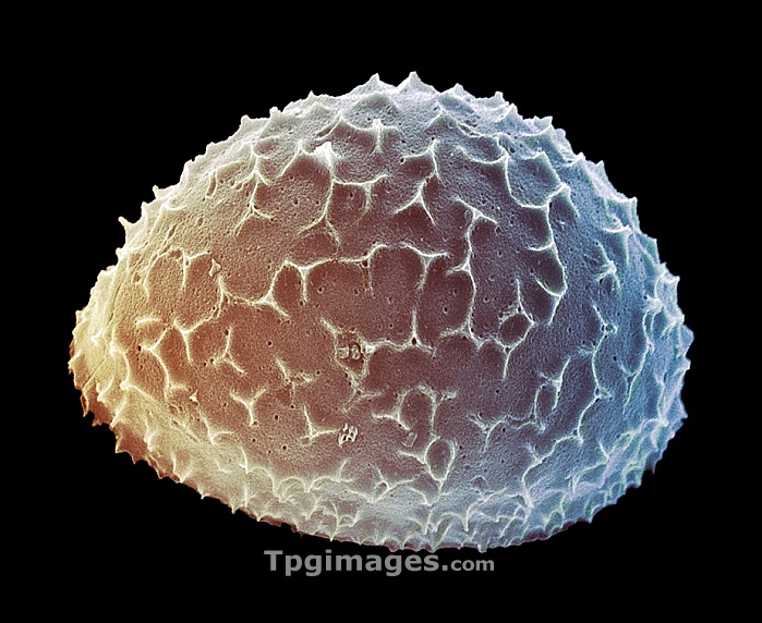
Diatom. Coloured scanning electron micrograph (SEM) of a Liradiscus sp. diatom. This is a planktonic unicellular alga. It has a mineralised cell wall (frustule) divided into two halves. The frustule contains silica and provides protection and support. Magnification: x95 at 6x7cm size.
- Image WebID: TPG05323488(RF)
- Same folder images: 20101126
- Available: immediately
- Comp : 2066X1691 (10.00 M)
Digital Size
px | px | dpi | = | cm | x | cm | = | MB |
Authorization Information
- Model Release:NO
- Property Release:NO
- Personality Rights: No
Copyright Information
Top Photo Corp. reserves the right to legally license the images and footage of this gallery, which must be licensed to Top Photo Corp. and shall be liable in full and liable Top Photo Corp. Damage (including but not limited to loss of business, loss of goodwill, litigation costs, attorneys' fees ...)
- diatom
- liradiscus sp.
- phytoplankton
- scanning electron micrograph
- sem
- coloured
- frustule
- radial symmetry
- symmetrical
- centric
- plankton
- planktonic
- nature
- botany
- plant
- alga
- algae
- algal
- diatoms
- cutout
- cutouts
- cut-out
- cut-outs
- cut out
- cut outs
- phytoplankton"
- phytoplankton"
- alga
- algae
- algal
- botany
- centric
- coloured
- cut out
- cut outs
- cut-out
- cut-outs
- cutout
- cutouts
- diatom
- diatoms
- frustule
- liradiscus sp.
- nature
- phytoplankton
- plankton
- planktonic
- plant
- radial symmetry
- scanning electron micrograph
- sem
- symmetrical

Similar Image
Similar Image>>