
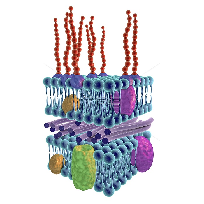
Gram-negative bacterial cell wall, illustration. The horizontal layers include both an external and an internal membrane (blue), both containing transmembrane proteins (green, yellow and purple). The membranes are separated by a thin peptidoglycan layer (purple rods). The outer surface of the external membrane is often a lipopolysaccharide layer with lipids (purple) in the membrane, and long saccharide side chains (red) extending out. This is termed a Gram-negative cell wall because it does not retain the Gram stain that helps identify microbial life.
| px | px | dpi | = | cm | x | cm | = | MB |
Details
Creative#:
TPG22478449
Source:
達志影像
Authorization Type:
RF
Release Information:
須由TPG 完整授權
Model Release:
N/A
Property Release:
N/A
Right to Privacy:
No
Same folder images:
artworkbacterialbacteriologybiochemicalbiochemistrybiologicalbiologycellstructurecellwallcellularcytologyexternalgramnegativegram-negativeillustrationinternallayerlayerslipidslipopolysaccharidemembranemembranesmicro-organismmicrobemicrobialmicrobiologypeptidoglycansaccharidessectionsectionedstructurestructures3d3dimensionalthreedimensionalwhitebackground
33dartworkbackgroundbacterialbacteriologybiochemicalbiochemistrybiologicalbiologycellcellcellularcytologydimensionaldimensionalexternalgramgram-negativeillustrationinternallayerlayerslipidslipopolysaccharidemembranemembranesmicro-organismmicrobemicrobialmicrobiologynegativepeptidoglycansaccharidessectionsectionedstructurestructurestructuresthreewallwhite
 Loading