
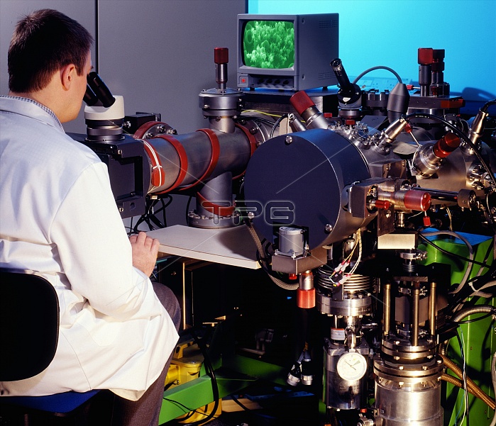
Scanning electron microscope. Researcher using a scanning electron microscope (SEM, upper left) to view blood cells. SEMs use an electron beam to obtain three-dimensional images of objects. The electron beam is moved across the sample, which causes secondary electrons to be emitted. It is these electrons which are used to form the image. SEMs can provide magnifications as high as 300,000 times original size. Photographed in the UK.
| px | px | dpi | = | cm | x | cm | = | MB |
Details
Creative#:
TOP10193920
Source:
達志影像
Authorization Type:
RM
Release Information:
須由TPG 完整授權
Model Release:
N/A
Property Release:
N/A
Right to Privacy:
No
Same folder images:
LANDSCAPESCIENCESCIENTIFICTECHNIQUETECHNIQUESTECHNOLOGYMICROSCOPYSCANNINGELECTRONMICROSCOPEMICROSCOPESSEMRESEARCHERSCIENTISTMANMALETECHNICIANUSINGWORKINGRESEARCHINGVIEWINGLOOKINGFOCUSSINGBLOODCELLCELLSREDTECHNOLOGYTECHNOLOGICALEQUIPMENTINSTRUMENTDEVICEMACHINEPHYSICSDISPLAYSCREENMONITORUNITEDKINGDOMUKHORIZONTALBRITAINBRITISH"
"BLOODBRITAINBRITISHCELLCELLSDEVICEDISPLAYELECTRONEQUIPMENTFOCUSSINGHORIZONTALINSTRUMENTKINGDOMLANDSCAPELOOKINGMACHINEMALEMANMICROSCOPEMICROSCOPESMICROSCOPYMONITORPHYSICSREDRESEARCHERRESEARCHINGSCANNINGSCIENCESCIENTIFICSCIENTISTSCREENSEMTECHNICIANTECHNIQUETECHNIQUESTECHNOLOGICALTECHNOLOGYTECHNOLOGYUKUNITEDUSINGVIEWINGWORKING
 Loading