
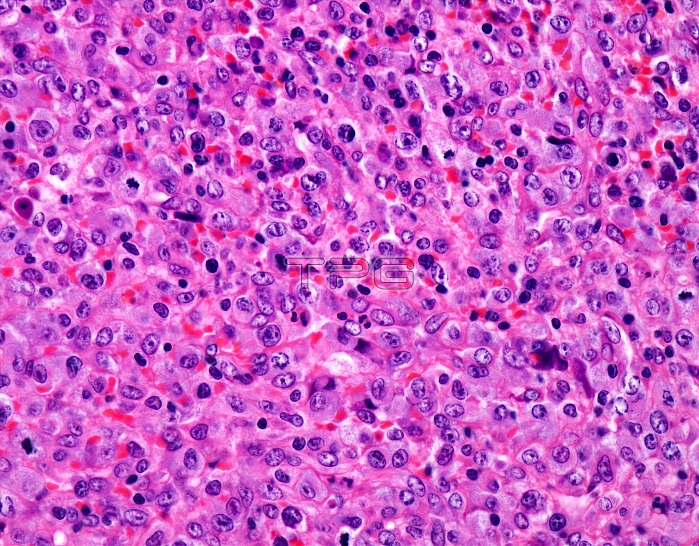
Peripheral T-cell lymphoma, light micrograph. In peripheral T-cell lymphoma, not otherwise specified, the tumour cells are variably-sized and consists of small cells with only minimal cytologic atypia and larger cells with irregular nuclear contours, prominent nucleoli, and brisk mitotic activity. Large cells with clear cytoplasm and Reed-Sternberg-like cells may be seen in some cases. Evidence of neoangiogenesis in the form of high endothelial venules is often seen. There is often a prominent component of reactive cells in the background, including small lymphocytes, eosinophils, macrophages, and plasma cells.
| px | px | dpi | = | cm | x | cm | = | MB |
Details
Creative#:
TOP24869239
Source:
達志影像
Authorization Type:
RM
Release Information:
須由TPG 完整授權
Model Release:
N/A
Property Release:
N/A
Right to Privacy:
No
Same folder images:
 Loading