
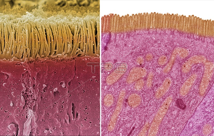
Intestinal microvilli. Comparison between a scanning election micrograph (SEM left) and transmission electron micrograph (TEM right) of an epithelial cell from a human small intestine, showing the densely packed microvilli. Each microvillus is approximately 1um long by 0.1um in diameter and contains a core of actin microfilaments. These tiny structures form a dense brush-like covering on the absorptive surfaces of the cells lining the small intestine. The cells absorb nutrients from digested food through the microvilli. Orange mitochondria are visible in the cytoplasm of the cell in the TEM image. Magnification: x3000 when printed at 10 centimetres wide. For a series of comparisons between SEMs and TEMs see images C047/7006 to C047/7034.
| px | px | dpi | = | cm | x | cm | = | MB |
Details
Creative#:
TOP25354337
Source:
達志影像
Authorization Type:
RM
Release Information:
須由TPG 完整授權
Model Release:
N/A
Property Release:
N/A
Right to Privacy:
No
Same folder images:
 Loading