
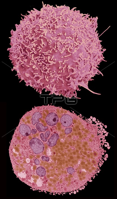
Comparison between a scanning election micrograph (SEM top) and transmission electron micrograph (TEM bottom) of a bladder cancer cell.Typical of many cancer cells the nucleus (purple TEM) is enlarged and multilobed, the cell walls are covered in projections (SEM) and the cytoplasm contains a large number of organelles (TEM) including brown lipid droplets. Most bladder cancers arise in the bladder lining. They may spread inwards or through the bladder wall into nearby organs, lymph glands and bones. Smokers and workers in the dye and rubber industries are at increased risk of the disease. It is also more common in tropical areas where the parasitic infection schistosomiasis is prevalent. Symptoms include blood in the urine and bladder infections. Treatment involves excision of affected tissues, with chemotherapy and radiotherapy. Magnification: x 3000 when printed at 10cm wide. For a series of comparisons between SEMs and TEMs see images C047/7006 to C047/7034.
| px | px | dpi | = | cm | x | cm | = | MB |
Details
Creative#:
TOP25354341
Source:
達志影像
Authorization Type:
RM
Release Information:
須由TPG 完整授權
Model Release:
N/A
Property Release:
N/A
Right to Privacy:
No
Same folder images:
 Loading