
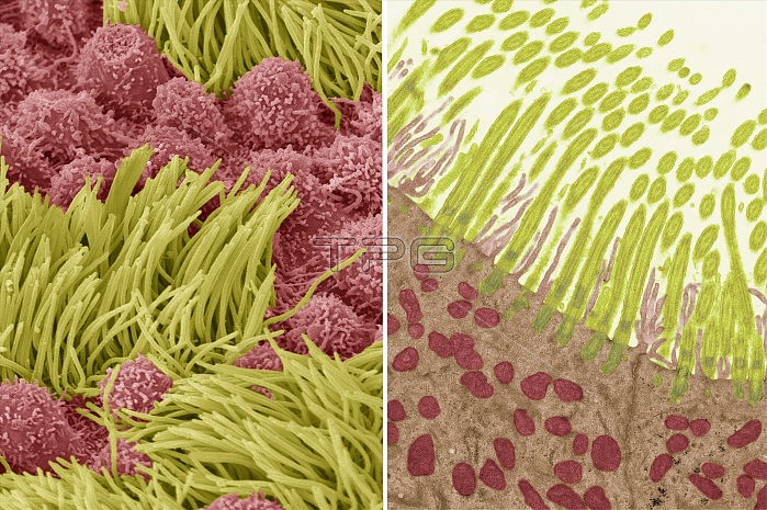
Comparison between a transmission election micrograph (SEM left) and scanning electron micrograph (TEM right) of tracheal epithelium. In both images cilia are coloured green while the microvilli of the mucous secreting goblet cells are red. In the TEM the internal structure of the cilia is visible; the mitochondria are seen in red within the cell. Mucous serves to trap tiny foreign particles in inhaled air which are transported by the movement of the cilia upwards and out of the respiratory tract, keeping the lungs and airways clear. In the SEM the surface structure of the specimen is visible. Magnification SEM X 5000 TEM X 9000 when printed at 10cm high. For a series of comparisons between SEMs and TEMs see images C047/7006 to C047/7034.
| px | px | dpi | = | cm | x | cm | = | MB |
Details
Creative#:
TOP25354346
Source:
達志影像
Authorization Type:
RM
Release Information:
須由TPG 完整授權
Model Release:
N/A
Property Release:
N/A
Right to Privacy:
No
Same folder images:
 Loading