
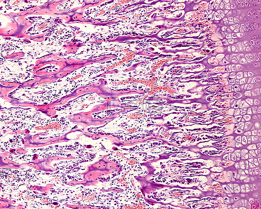
Light micrograph of the epiphyseal growth plate of a developing long bone. The epiphyseal cartilage shows the following layers, from right to left: zone of proliferation, zone of hypertrophy with large lacunae, zone of calcification (not visible as such, but can be identified by the degeneration of the chondrocytes leaving empty cartilaginous lacunae), and an ossification zone, where there is an invasion of blood vessels and osteogenic cells. The mixed trabeculae with a bluish centre of acellular calcified cartilage, covered by pink primary bone tissue with osteocytes can be seen further left.
| px | px | dpi | = | cm | x | cm | = | MB |
Details
Creative#:
TOP25928991
Source:
達志影像
Authorization Type:
RM
Release Information:
須由TPG 完整授權
Model Release:
N/A
Property Release:
N/A
Right to Privacy:
No
Same folder images:
 Loading