
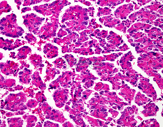
Choroid plexus carcinoma, light micrograph. Choroid plexus tumours comprise爈ess than 1% of all brain tumours. Almost?0% of cases occur in children爉aking up 2% to 4% of pediatric brain tumours. The most common location is爈ateral ventricles. Given their location, the presenting symptoms and signs are related to爃ydrocephalus燼nd爄ncreased intracranial pressure. Choroid plexus carcinomas are爏olid, infiltrative tumours爁requently with foci of爃aemorrhage and necrosis. Choroid plexus carcinomas show papillary architecture in better differentiated foci (as shown in this image). Poorly-differentiated areas are generally solid and composed of highly atypical cells arranged in sheets. The papillary cores in this choroid plexus carcinoma are lined by anaplastic cells with irregular hyperchromatic nuclei.
| px | px | dpi | = | cm | x | cm | = | MB |
Details
Creative#:
TOP25929018
Source:
達志影像
Authorization Type:
RM
Release Information:
須由TPG 完整授權
Model Release:
N/A
Property Release:
N/A
Right to Privacy:
No
Same folder images:
 Loading